Cross Section Of A Compact Bone : File Compact Bone Decalcified Cross Section Jpg Wikimedia Commons / Compact bone (cross section of dried bone).. Cross section of the compact bone. (micrograph provided by the regents of university of michigan. Bone decalcification is the removal of the mineral component using an acid, leaving the bone soft and easy to cut. Hope you enjoy and please. Most bones contain both compact and spongy bone.
These are abundant and characteristic of compact bone. Select different colors for the. The innermost layer of membrane is made up of. The outlined area is a cross section of an osteon of compact bone. The two layers of compact bone and the interior spongy bone work together to protect the internal organs.
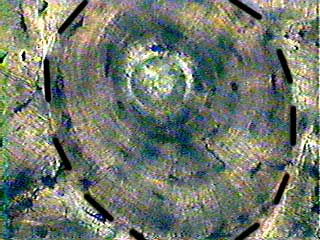
Transverse Section Of Compact Bone Clipart Etc from etc.usf.edu This is a short tutorial using blender 2.8 that shows how to create a bone cross section and using images to create the textures. Also called cortical bone, the compact variety usually features a haversian system, or cylindrical unit within the structure. Canaliculi allow the passage of interstitial fluid between the central canal and the lacunae housing osteocytes. As seen in the image below each osteon is also composed of a number of different cells responsible for the maintenance of the bones, including osteocytes and osteoblasts. (b) in this micrograph of the osteon, you can clearly see the concentric lamellae and central canals. Observe that the matrix of the bone is deposited in concentric layers that are called lamellae (5). From wikimedia commons, the free media repository. The spongy and compact bone tissue in the cross section of a skull bone.
Jump to navigation jump to search.
Concentric layers of bone cells (osteocytes). Dry bone is cut and polished before mounting on a slide. This model shows a cross section of compact bone. A central tube called a haversian canal typically runs in the same path as the length of the bone. Compact bone, dense bone in which the bony matrix is solidly filled with organic ground substance and inorganic salts, leaving only tiny spaces that contain the osteocytes, or bone cells. (micrograph provided by the regents of university of michigan. Structures and bone areas in column b, and use them to color the coding. Bone decalcification is the removal of the mineral component using an acid, leaving the bone soft and easy to cut. In three dimensions an osteon is cylindrical in shape. Compact bone is very different from the other tissues you have seen. Also called cortical bone, the compact variety usually features a haversian system, or cylindrical unit within the structure. In the last decade, considerable technological improvements have been made to repair damaged bones and tissue, such as bone cross sections with implants for microscopic examinations. They build the entire picture, improve your understanding, consolidate the information and facilitate recall.
Jump to navigation jump to search. This model shows a cross section of compact bone. As the names suggest compact bone looks compact and the spongy bone looks like sponges. An estimated 10 percent of an adult's skeleton is replaced each year. In three dimensions an osteon is cylindrical in shape.
Bone Compact Ground C S from www.austincc.edu This is a cross section through decalcified bone. As the names suggest compact bone looks compact and the spongy bone looks like sponges. Jump to navigation jump to search. Most but not all human bones have circular cross sectional shapes. Concentric layers of bone cells (osteocytes). Cross section of compact bone. Also called cortical bone, the compact variety usually features a haversian system, or cylindrical unit within the structure. A central tube called a haversian canal typically runs in the same path as the length of the bone.
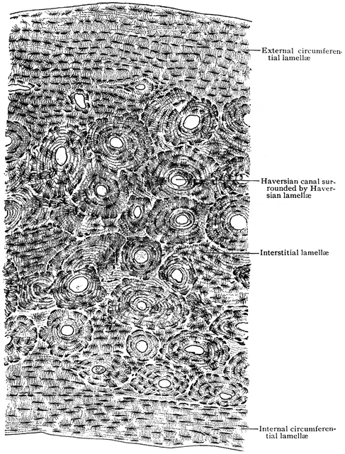
A cross section of a compact bone shows concentric circles called lamellae.
Compact bone, dense bone in which the bony matrix is solidly filled with organic ground substance and inorganic salts, leaving only tiny spaces that contain the osteocytes, or bone cells. In three dimensions an osteon is cylindrical in shape. A cross section of a compact bone shows concentric circles called lamellae. The spongy and compact bone tissue in the cross section of a skull bone. Canaliculi allow the passage of interstitial fluid between the central canal and the lacunae housing osteocytes. There are two ways to study bone histology. Dry bone is cut and polished before mounting on a slide. Compact bones make up 80 percent of the human skeleton; In the center of each osteon is the central canal, a space that houses blood vessels and nerves that supply bone. Select different colors for the. (micrograph provided by the regents of university of michigan. Their course follows the main axis of long bone. The two layers of compact bone and the interior spongy bone work together to protect the internal organs.
These are mostly compacted bone with little marrow and include most of the bones in the limbs. Observe that the matrix of the bone is deposited in concentric layers that are called lamellae (5). Most bones contain both compact and spongy bone. The outlined area is a cross section of an osteon of compact bone. There are two ways to study bone histology.
Bone Canaliculus Wikipedia from upload.wikimedia.org The outlined area is a cross section of an osteon of compact bone. This model shows a cross section of compact bone. Compact bone (cross section of dried bone). Their course follows the main axis of long bone. I am sure they have a higher strength vs weight ratio. The innermost layer of membrane is made up of. Jump to navigation jump to search. These are abundant and characteristic of compact bone.
Compact bone is very different from the other tissues you have seen.
There are two ways to study bone histology. This model shows a cross section of compact bone. The remaining material is mostly collagen. These are abundant and characteristic of compact bone. Cross section of compact bone. Magnification view of compact bone tissue. Also called cortical bone, the compact variety usually features a haversian system, or cylindrical unit within the structure. The innermost layer of membrane is made up of. The basic unit of structure in this type of bone is the haversian system, or osteon. The connection point for the periosteum. In the center of each osteon is the central canal, a space that houses blood vessels and nerves that supply bone. These are mostly compacted bone with little marrow and include most of the bones in the limbs. Osteocyte processes lie in tiny canals (canaliculi) in the bone matrix.
A cross section of a compact bone shows concentric circles called lamellae cross section of a bone. There are trabeculae in spongy bone which gives its sponge like appearance.


0 Komentar